9. The heart and the circulatory system
Contents of the chapter
9.1 Discover the circulatory system
9.2 The cardiovascular system
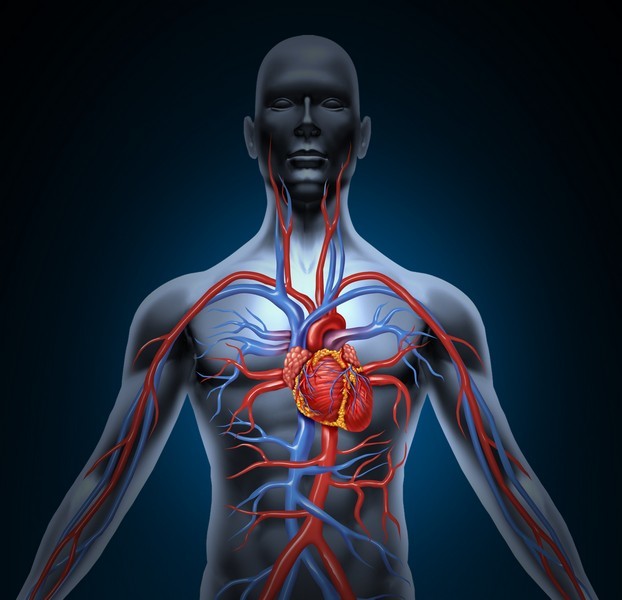
The circulatory or cardiovascular system is made up of three main parts - the heart, the blood vessels and the blood that flows through them.
The heart is a hollow muscle located inside the thoracic cavity, behind the sternum. It is protected by the ribs of the chest. An adult heart weighs about 300g and is the size of its owner's fist.
The function of the heart is to pump blood to different parts of the body. The heart is divided into two halves, the right and the left. The septum separates the right-hand and left-hand sides of the heart. Both halves are divided into atria and ventricles. The atria (plural of atrium) are where the blood collects when it enters the heart. As the heart contracts, the blood leaves the ventricles through the arteries and is take returns to the atria via the veins.
The heart does its work tirelessly. This is made possible by the fact that it rests briefly between its beats. This periodicity of contractions and pauses is called the heart rate. The heart rate can be measured from the blood vessels of the wrist or by using a blood pressure monitor.
The blood pumped by the heart circulates into the body via the blood vessels. They include thick-walled arteries. The main artery is called aorta. It also includes thin-walled and narrow capillaries, as well as veins.
The heart functions independently. However, when necessary, the function of the heart can be influenced by both the nervous system and hormones.
9.3 The structure of the heart
The heart’s four chambers function as a double-sided pump. The heart needs to work at just the right rhythm. It is divided into right and left halves. These halves are further divided into atria and ventricles. The right ventricle pumps blood to the lungs whereas the left ventricle pumps blood to everywhere else in the body. For this reason, the left ventricle has the thickest muscle mass of all the chambers.
Veins carry blood both to the right and left atria at the same time. The cycle begins when the two atria contract, which pushes blood into the ventricles. After this, the ventricles contract, which forces the blood out of the heart.
The valves between the atria and the ventricles prevent blood from returning to the ventricles. When the heart is resting between its beats, the valves between the ventricles and arteries prevent the blood from flowing back into the heart. However, there are no valves between the atria and the veins.
9.4 The function of the heart
The rhythm of heartbeat is maintained by the sinoatrial (SA) node. The impulse starts in a small bundle of specialized cells located in the right atrium. The electrical activity spreads through the walls of the atria and causes them to contract. This forces blood into the ventricles.
The heart beats at an average rate of 60 to 75 beats per minute and pumps about five litres of blood. This means that the total volume of a person's blood is moved in a period of just one minute. Under intense exercise, your heart rate can increase by more than two hundred beats.
Thus, the heart can beat up to 100,000 times each day. During this time, the heart will pump almost 7,000 litres of blood. The heart can do this because it can rest for a combined time of four hours between beats each day.
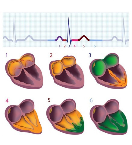
The image shows the electrical activity of the heart (ECG).
9.5 Coronary arteries
 Like all other muscles, the heart needs oxygen and nutrients in order to function properly.
Coronary arteries branch from the main artery, the aorta. They supply the heart muscle with oxygen.
The condition of the coronary arteries is crucial for the functioning of the heart. Their congestion and calcification can lead to a lack of oxygen in the heart. Ultimately, these processes can lead to a heart attack.
Coronary artery diseases are caused by the narrowing or blockage of the coronary arteries. This restricts the supply of oxygen to the heart.
9.6 Pulmonary and systemic circulatory system
In order to understand the circulatory system, it is good to remember the following three things:
- The heart has two halves: right and left. There is no movement of blood between them.
- Types of blood vessels: arteries carry blood from the heart, and veins carry blood to the heart. The capillaries are small vessels between the cells and the larger blood vessels.
- During one heartbeat and the following resting period, all of the following things happen:
As the heart rests, the inferior vena cava and the superior vena cava carry carbon dioxide-rich blood from all over the body to the right atrium. At the same time, oxygen-rich blood is carried from the lungs to the left atrium. From the atria, blood flows immediately to the right and left ventricles.
Pulmonary arteries carry the blood into the lungs where carbon dioxide is replaced by oxygen. This takes place in the pulmonary capillaries, where carbon dioxide is removed from the blood and oxygen is bound to red blood cells. This oxidized blood returns to the heart via the pulmonary veins.
From the left ventricle, the blood, which is now under a high pressure, exits the heart via the aorta. The arteries that branch from the aorta carry the blood everywhere in the body. Nutrients and oxygen are released to all the cells of the body through the thin walls of capillaries.
The blood flow from the left ventricle of the heart to the right atrium is called systemic circulation. The circulation of blood in the lungs (right ventricle → lungs → left atrium) is called pulmonary circulation.
Image: A = left atrium, B = left ventricle, C = right atrium, D = right ventricle, E = lungs, F = organs.
1 = aorta (artery), 2 = artery, 3 = capillaries, 4 = vein, 5 = inferior vena cava, 6 = superior vena cava, 7 = pulmonary artery, 8 = pulmonary vein, 9 = lymphatic vessel and lymph node.
Cellular respiration uses the oxygen transported by the blood and produces toxic carbon dioxide. The inferior and superior vena cava bring oxygen-poor blood from the body into the right atrium.
9.7 Blood pressure
Arterial blood pressure is expressed as a measurement with two numbers. In this measurement, one number is found on the top (systolic) and another is located on the bottom (diastolic), like a fraction. The top number refers to the amount of pressure in your arteries during the contraction of your heart muscle, whereas the bottom number refers to your blood pressure when your heart muscle is resting between beats. The average reading for a young adult is 120/80 mmHg.
In hypertension (high blood pressure), the blood pressure readings are significantly higher than normal. Symptoms of hypertension may include headache, dizziness and visual disturbances. Maintaining a healthy diet and exercising are good methods of preventing cardiovascular disease.
If the venous valves in the lower limbs are not working properly, blood can flow back into the veins and the pressure inside the veins can increase. This results in varicose veins, as the fluid is drained from the veins to the surrounding tissues. Typically, varicose veins can be found just under the skin.
9.8 Electrocardiography (ECG)
An electrocardiogram (electro refers to electricity, cardio to the heart and gram to a curve) is used to determine the stages of heart contraction.
Take a look at the image and the colored middle part of the curve with numbers 1-6. There is electrical activity in the yellow-coloured parts of the heart.
In steps 1 and 2, the electrical impulses occur in the atria. As a result, the atria contract and the blood flows to the ventricles.
The electrical impulse is transmitted to the muscles of the ventricular walls. The ventricles contract (step 3), and the blood flows into the aorta and pulmonary arteries.
In step 5, the ventricles relax. In step 6, all muscle cells inside the heart are at rest.
9.9 Blood vessels
Arteries are the blood vessels that transport blood away from the heart. When the ventricles of the heart contract, blood is transported into the arteries. Arteries have thick muscular walls, which help them withstand high pressures caused by blood leaving the heart.
The largest artery is the aorta, which begins from the heart's left ventricle. The aorta branches above the heart into several arteries, which continue to branch into thousands of smaller arterioles throughout the body.
Arterioles connect with even smaller blood vessels called capillaries. You can see these capillaries, for example, in the white of your eyes. Oxygen and nutrients are exuded through the walls of the capillaries into the body's cells.
All harmful substances produced in the body are transported from the intercellular space back to the capillaries for further transport.
Veins are the blood vessels that transport blood to the heart. They carry oxygen-poor blood, and therefore appear as blue through the skin. Little pressure remains by the time blood leaves the capillaries and enters the venules. The flow of blood back to the heart is referred to as venous return. It is dependent on muscle action.
The muscle contractions near the veins squeeze the blood through the valves. The venous valves prevent blood from flowing backwards.

9.10 The lymphatic system
The lymphatic system consists of lymphatic vessels and lymph nodes. Blood plasma leaks into the body's tissues through the thin walls of the capillaries. The lymphatic system functions to remove these fluids and materials from tissues, returning them to the bloodstream via lymphatic vessels. The lymphatic system plays a key role in transporting fats from the intestines.
The structure of the lymphatic vessels resembles that of the blood vessels, as the lymphatic vessels also have valves that causes lymph to eventually flow forward instead of travelling backwards. The contraction of your muscles and the pressure in the arteries becomes the pump that helps the fluid move around your body. Exercise can help the lymphatic system to function more effectively.
The lymph passes through lymph nodes before returning to the bloodstream via veins located near the heart. Lymph nodes can be located, for example, in the armpits and the groin. They are rich in white blood cells that help fight microbes that may have entered your lymphatic system. If you have a severe flu, your doctor may test your neck to see if your lymph nodes are swollen.

Left: The lymphatic vessel collects tissue fluid from between cells. Right: The lymphatic system.


