14. Eyesight
Contents of the chapter
14.1 The eye
Eyesight is one of our most important senses. The eyes are located deep under the protection of the skull. Eyelashes also protect our eyes. Tear fluid cleanses the cornea.
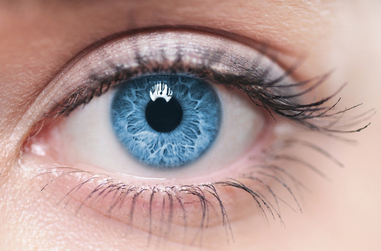
The pupil (the middle of the lens) as well as the iris and the transparent cornea are the visible parts of the eye. The rest of the eye is located inside the skull.

14.2 Human eyesight
 Human eyesight is characterized by eye coordination, which is the ability of both eyes to work together as a team. Each of your eyes sees a slightly different image. Your brain blends these two images into one three-dimensional picture through a process called fusion. This makes us able to better estimate things like distances between objects. This binocular vision is typical of primates and predators, for whom accurate estimation of distances is necessary for survival.
Even a weaker ability to estimate distances is sufficient for a rabbit, as long as it can be used to distinguish movement from the largest possible area. The rabbit finds its food by pressing its head down. The rabbit's eyes are on different sides of its head, which means that it has no binocular vision.
14.3 The anatomy of the human eye
 The human eye is protected by a light-transmitting and refracting cornea (2).
The iris (4) is located beneath the cornea. The iris regulates the size of the pupil. The colour of the iris varies from brown to light grey. Only a percentually small segment of people have blue irises.
The lens (1) is located under the iris. The lens refracts light strongly. The ciliary muscle (5), part of the ciliary body, controls the shape of the lens.
The pupil (3) is located at the center of the lens. It is the opening in the middle of the iris.
The vitreous body (6) consists of a gel-like liquid that fills the eye.
The retina (7) is found at the very back of the eye. Light-sensing neurons are located on its surface. The retinal nerve cells consume a lot of energy and oxygen, which is why there is a choroid behind the retina, the tiny capillaries of which bring sugar and oxygen into place.
The axons of the retinal neurons form the optic nerve (8). One optic nerve connects each eye to the brain. The choroid is the vascular layer of the eye that lies between the retina and the sclera.
Each of our eyes has a tiny functional blind spot about the size of a pinhead. In this tiny area, where the optic nerve passes through the surface of the retina, there are no light-detecting photoreceptor cells. Since there are no photoreceptor cells in this spot, the result is a blind spot. Without photoreceptor cells, the eye cannot send any messages about the image to the brain, which usually interprets the image for us. However, the brain connects information from both eyes, which is why we don’t notice the “blind spot” in our retinas.
14.4 Optic nerves
The light-sensitive layer of cells at the back of the eyeball is called the retina. It is the equivalent of the film in a camera. This layer is comprised of two types of cells: rods and cones.
People are able to see very different colours. With the help of colour vision, our ancestors were able to identify ripe fruit from raw. Indeed, our sense of sight has partially replaced our weakened sense of smell.
The cone cells are photoreceptor cells in the retinas that specialize in colour separation. They are most abundant in the middle of the retina, in the yellow spot. The cones require a large amount light to operate and are functional during daylight but are almost useless at night.
 Fundus imaging of the retina covers the back of the eye. The image shows the main structures of the fundus: the retina, the optic nerve head, the area of accurate vision, and the main blood vessels.
The picture below shows red and green chilies. Some people are unable to distinguish between red and green. This condition is known as color blindness. It is more common in men than in women.
 Do you distinguish between the red and the green chilies?
When we want to focus our gaze on something very small or distant, we turn our eyes so that the light is directed directly at the yellow spot. This increases the resolution of the image produced. However, not all people see colours the same way. The colour separation ability can vary greatly. The most typical is red-green colour blindness, which makes it difficult to distinguish red tones from green ones. Some people do not see colours at all.
The rod cells incorporate rhodopsin, sometimes called “visual purple,” a light-sensitive protein that activates the rod cells and allows you to see at night. Night vision does not start forming immediately when the lights go out, so it takes a while to adapt to twilight.
Visual purple decomposes rapidly under the influence of light, so for example, turning on the lights in complete darkness can cause a moment of glare. Reading in the dark or very close does not impair vision, but can strain the muscles of the eyes. This can cause headaches, for example. Moving a book or display terminal a little further or resting your eyes by looking far in between is useful.
14.5 Eye function

The cornea located at the front of the eye refracts light onto the lens. The lens, on the other hand, refracts light precisely on the surface of the retina. The shape of the lens can be changed by contracting the ciliary muscles, which makes us able to focus our gaze near or far.
- When focusing on a distant object the ciliary muscles are at rest.
- When focusing on a nearby object the ciliary muscles contract.
The image illustrates the contraction of the ciliary muscle when looking at a distant or a nearby object. The parts of the eye: 1. ciliary muscle, 2. pupil, 3. lens.
When we focus on an object in a distance (upper image), the ciliary muscles are at rest, suspensory ligaments are stretched, and the lens is thin. Light is refracted only slightly.
When we look at something up close (lower image), the ciliary muscles are contracted, the suspensory ligaments loosen, and the lens becomes thicker, more convex. Light is refracted strongly. With age, the convexity of the lens decreases.
14.6 Refractive errors
 The shape of the eye and the thickness of the cornea can vary between different people. If the cornea is irregular, light rays enter the eye at different angles, and do not focus on one point the retina, but on many different points. This results in a blurred, distorted image.
Adolescents can develop nearsightedness or myopia. Myopia is a common vision condition which occurs when the shape of the eye causes light rays to bend (refract) incorrectly. A myopic person can see objects near to them clearly, but the objects farther away seem blurry. Nearsightedness can be corrected with glasses that have concave lenses or through refractive surgery.
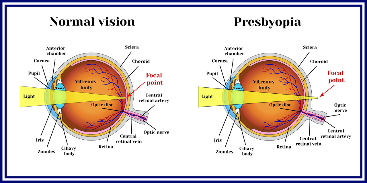
As we age, muscles that control our pupil size and reaction to light lose some strength. This causes the rays of light to refract behind the retina. In this case, the person sees very far, but the near vision is impaired. Long-distance refraction can be corrected with glasses that have convex lenses.
Far-sightedness, presbyopia, is caused by loss of elasticity of the lens of the eye. It typically develops in middle age, when the person is at the age of 40-45 years.


