18. From fertilization to birth
Contents of the chapter
18.1 Gamete formation and fertilization
 In males, gamete production takes place in the testes. The male gamete is called sperm. Female gametes, called ova, are produced in the ovaries.
In men, sperm cells are produced all the time. In females, a single ovum or egg cell matures about once a month.
Gametes carry half of an individual's genetic information (23 chromosomes). Gametes are created in meiosis.
In germ cell fusion or fertilization, which occurs as a result of sexual intercourse, the male and female gametes are fused together. By doing so, they form a fertilized egg cell. This new cell is diploid, which means that it has 46 chromosomes. Half of these chromosomes come from the mother, whereas the other half come from the father.
The fertilized egg cell marks the beginning of a human's individual development. A new human individual begins to develop inside the mother's womb.
18.2 Fertilization
In humans, pregnancy is possible when sexual intercourse occurs at the same time as the female is ovulating. After ovulation, the egg lives for 12 to 24 hours. The egg must be fertilized in that time for a woman to become pregnant.
Human sperm will remain fertile for 1–2 days. When the sperm arrives in the acidic conditions of the vagina, they swim forward towards the uterus and the fallopian tube, where fertilization takes place.
Out of the strongest and fastest sperm, only one sperm cell penetrates the surface of the ovum. Fertilization occurs when the nuclei of a sperm cell and an egg cell fuse together. When this happens, the two gametes form a diploid cell, known as a zygote. The ovarian membrane becomes impermeable to sperm immediately after fertilization.

Fertilization: 1. The sperm cell. 2–3. The acrosome reaction makes it possible for the sperm cell to penetrate the zona pellucida. The cell membranes of the sperm cell and the egg cell are combined. 4. The nucleus of the sperm cell moves inside the egg.
18.3 Embryonic development
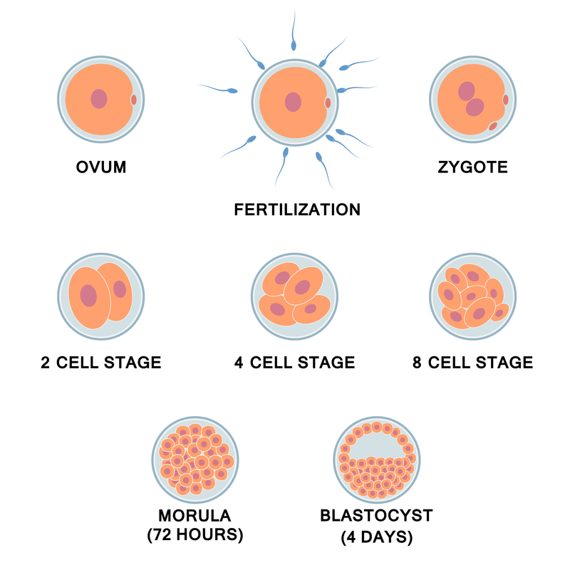
 After fertilization, the newly formed single-celled zygote begins to divide into multiple cells. The number of cells doubles during each division.
About a week after fertilization, a hollow ball of cells called a blastocyst arrives in the uterus. The blastocyst is then implanted on the wall of the uterus.
At this point, the rapidly developing embryo is still not larger in size than the fertilized egg.
The zygote initially receives nutrients from the ovum. Once attached to the uterus, the embryo receives nutrients from a yolk sac until the umbilical cord and placenta have developed.
In an eight-week-old embryo, all the major organs have already been developed. From this point onwards, the developing individual is called a fetus. The eight-week-old fetus is approximately three centimeters long, and its head and limbs can already be distinguished. Its faint heartbeat is also already noticeable.
Image on the right: The first stages of human embryonic development.
18.4 The fetus in the uterus
The placenta is an organ that provides oxygen and nutrients to the fetus and removes waste products from it. The placenta attaches to the wall of the uterus, and the baby's umbilical cord arises from it.
 The fetus in the uterus.
The fetus receives oxygen, nutrients, hormones and antibodies from the mother's bloodstream. Water and harmful substances such as urine and carbon dioxide are released from the fetus into the mother's blood. The placenta filters out most harmful substances, but is permeable to alcohol, tobacco toxins, viruses, bacteria, certain drugs and narcotics. This is why consuming such products during pregnancy is harmful to the fetus.
18.5 Fetal development
The fetus develops quickly during the first eight weeks of pregnancy. All the major organs and structures of the body are formed during this time. After this, the development will continue mainly as growth. From now on, the individual's susceptibility to various developmental disturbances and malformations decreases. This reduces the chance of miscarriage. Inside the uterus, the fetus moves in amniotic fluid, while the amniotic sac and the wall of the uterus provide it with protection.
 The stages of fetal development.
After the fifth month of pregnancy, the fetus is approximately 30 centimeters long and weighs nearly 500 grams.
As the nervous system develops, the growing fetus begins to receive information from the outside world through various senses. For example, the sense of touch manifests itself in response to the mother’s movements. Similarly, thanks to the development of the sense of hearing, the mother’s voice becomes familiar to the child even before birth.
During the last months of pregnancy, a layer of fat forms under the skin of the fetus. This helps the fetus tolerate the lower temperature of its environment after birth.
At the beginning of the 35th week of pregnancy, the fetus is usually settled in the uterus so that it is ready to be born headfirst. If the fetus is in a different position, a caesarean section is often carried out.
A human pregnancy lasts for approximately nine months, or 40 weeks. During this time, the fetus grows to approximately 50 centimetres in length, and weighs about three kilograms. In a 38-week-old fetus, the individual's lungs are already developed so that no attempt is made to slow or prevent the course of labor if there are signs that it is starting.
Changes also occur in the mother's body during fetal development. Potential fatigue and nausea often pass after early pregnancy. Maternal weight increases slowly at first, but grows rapidly after mid-pregnancy, due to weight of the fetus, amniotic fluid and placenta, as well as an increase in blood and other body fluids. On average, a pregnant woman experiences a weight gain of approximately ten kilograms, about half of which is lost during childbirth. Pregnant women should regularly visit a clinic, where the well-being of both the mother and the fetus are monitored.
Fetal development can be examined via ultrasound imaging or by taking an amniotic fluid sample or a maternal blood sample. At the clinic, the weeks of pregnancy are calculated from the beginning of the last menstrual period. From there, embryonic and fetal development takes 40 weeks until birth. Because ovulation occurs about 14 days after the onset of menstrual bleeding, the actual duration of pregnancy is 38 weeks.
18.6 End of pregnancy and childbirth
There are changes in the mother's condition during the last weeks of pregnancy. As the head of the fetus settles into the opening formed by the bones of the mother’s pelvis and the base of the uterus descends lower, the mother may again feel easier to breathe when the upper part of the uterus does not press on the lungs. Instead, the uterus presses on the bladder.
The actual initiator of labor is unknown, and therefore its exact time cannot be known in advance. There are several factors that are thought to affect it, including changes in hormone levels. The proportion of progesterone, which maintains pregnancy, in the mother's blood decreases, whereas the proportion of oxytocin, which promotes contractions, increases.
Childbirth progresses in three stages. The first stage starts with contractions and the cervix dilating, and ends when the cervix is fully open.
Next, during the rather short second or effort stage, the uterine muscles begin their work. During this stage, the mother must actively push the baby out through the birth canal. In the third stage, the mother pushes out a placenta shaped like a small frying pan, as well as the rest of the umbilical cord and the fetal membranes.
Human labor pains are more severe compared to those experienced by other mammals. In an upright human, the pelvic orifice is narrower than in mammals that walk on four limbs. In addition, the head of a human baby is large when compared to the rest of its body.
The fetus does not always place itself head-down in the mother's womb during late pregnancy. If the attempt to turn is unsuccessful, a caesarean section is carried out. In some cases, the mother gives birth to the child feet first. The C-section can also be carried out, for example, due to a strong fear of childbirth on the mother's part, or when the size of the mother's pelvic opening is too small for the fetus. During the childbirth phase, an emergency C-section may need to be carried out if the mother's fetal heart rate falls. The mother's abdomen is cut open, and the child is lifted from the uterus. The uterus and abdominal layers are then sutured closed. A mother who gives birth without a C-section usually recovers from childbirth more quickly, simply because she does not need to be wary of a healing sectional wound.
After giving birth, the mother's abdomen is still the size of a pregnant belly. This happens because the uterus takes 4 to 8 weeks to contract back to its normal size after the fetus is removed.
After birth, the newborn child has to get used to many changes in its environment. This process begins when the baby takes its first breath, filling its lungs with air as breathing begins. The child's blood circulation begins, and oxygenated blood now comes from the lungs instead of the umbilical cord. The baby needs to learn to be fed. Being close to its mother during feeding helps the baby to get used to the coldness, light and sounds of the surrounding world.

18.7 Twins
Sometimes, there might be two ova in the female's fallopian tube. When both of these ova are fertilized, two fetuses can begin to develop in the uterus at the same time. In this case, the twins are called dizygotic ('non-identical') or 'fraternal'.
Twins can also develop from one zygote, which divides into two embryos. These twins are called monozygotic ('identical'), meaning that they develop from one zygote, which splits and forms two embryos, Children have exactly the same genome and are called identical twins (twins with the same egg).

Dizygotic twins and monozygotic or identical twins.

